- Home
- Medical news & Guidelines
- Anesthesiology
- Cardiology and CTVS
- Critical Care
- Dentistry
- Dermatology
- Diabetes and Endocrinology
- ENT
- Gastroenterology
- Medicine
- Nephrology
- Neurology
- Obstretics-Gynaecology
- Oncology
- Ophthalmology
- Orthopaedics
- Pediatrics-Neonatology
- Psychiatry
- Pulmonology
- Radiology
- Surgery
- Urology
- Laboratory Medicine
- Diet
- Nursing
- Paramedical
- Physiotherapy
- Health news
- AYUSH
- State News
- Andaman and Nicobar Islands
- Andhra Pradesh
- Arunachal Pradesh
- Assam
- Bihar
- Chandigarh
- Chattisgarh
- Dadra and Nagar Haveli
- Daman and Diu
- Delhi
- Goa
- Gujarat
- Haryana
- Himachal Pradesh
- Jammu & Kashmir
- Jharkhand
- Karnataka
- Kerala
- Ladakh
- Lakshadweep
- Madhya Pradesh
- Maharashtra
- Manipur
- Meghalaya
- Mizoram
- Nagaland
- Odisha
- Puducherry
- Punjab
- Rajasthan
- Sikkim
- Tamil Nadu
- Telangana
- Tripura
- Uttar Pradesh
- Uttrakhand
- West Bengal
- Medical Education
- Industry
Increased extraocular muscle volume tied to dysthyroid optic neuropathy in patients with thyroid eye disease

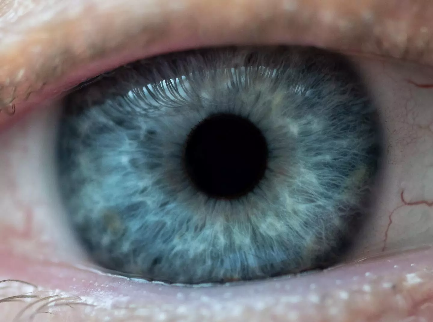
CAPTION
Researchers have developed a potential new treatment for the eye disease glaucoma that could replace daily eye drops and surgery with a twice-a-year injection to control the buildup of pressure in the eye.
CREDIT
Rob Felt, Georgia Tech
Increase in extraocular muscle volume tied to dysthyroid optic neuropathy in patients with thyroid eye disease suggests a new study published in the Canadian Journal of Ophthalmology. A study investigated extraocular muscle volumes in thyroid eye disease (TED) patients with and without dysthyroid optic neuropathy (DON). The extraocular muscles were manually segmented into consecutive axial and coronal slices, and the volume was calculated by summing the areas in each slice and multiplying by the slice thickness. Data were collected on patient demographics, disease presentation, thyroid function tests, and antibody levels. Results: Imaging from 200 orbits was evaluated. The medial rectus, lateral rectus, superior muscle group, inferior rectus, and superior oblique volumes were significantly greater in orbits with dysthyroid optic neuropathy than TED orbits without dysthyroid optic neuropathy (p < 0.01 for all). There was no significant difference in the inferior oblique muscle volume (p = 0.19). An increase in the volume of the superior oblique muscle showed the highest odds for dysthyroid optic neuropathy. Each 100 m3 increase in superior oblique, lateral rectus, inferior rectus, medial rectus, and superior muscle group volume was associated with 1.58, 1.25, 1.20, 1.16, and 1.14 times increased odds of dysthyroid optic neuropathy. All extraocular muscle volumes except for the inferior oblique were significantly greater in dysthyroid optic neuropathy patients. Superior oblique enlargement was associated with the highest odds of dysthyroid optic neuropathy, suggesting superior oblique enlargement to be a novel marker of dysthyroid optic neuropathy.
Dr. Shravani Dali has completed her BDS from Pravara institute of medical sciences, loni. Following which she extensively worked in the healthcare sector for 2+ years. She has been actively involved in writing blogs in field of health and wellness. Currently she is pursuing her Masters of public health-health administration from Tata institute of social sciences. She can be contacted at editorial@medicaldialogues.in.


